
Coupled with the output of Fusion MR Viewer, Sonablate® HIFU is changing the standards for prostate cancer treatment. Together the two work to deliver minimally invasive, targeted focal therapy. Sonablate HIFU uses high intensity focused ultrasound (HIFU) waves to ablate lesions without damaging surrounding tissue. It delivers the HIFU treatment, but requires an MRI-US fusion biopsy platform, like the Fusion Bx, to visualize and target lesions. As a standalone system, procedures are limited to whole gland or hemi-gland (half of the prostate) ablations. Without fusion, the procedure relies on ultrasound guidance to identify lesions, negating the key benefit of focal therapy.
How Does Sonablate HIFU Work?
Typically, the probe is supported by an articulated probe holder (or stepper) accessory attached to the operating table. Encased by a balloon-like sheath, the probe contains two transducers, of different focal lengths, which both image and deliver the HIFU treatment. Once emitted, the high intensity ultrasound waves travel through the rectal wall and converge at a single focal point to heat and destroy targeted cancerous cells. Since the ultrasound waves are individually weak, the treatment spares critical areas and surrounding tissue from harm. To cool the rectal wall and HIFU transducer, the chiller circulates cold water to the tip of the probe through the balloon. As the volume of water is adjusted, the balloon becomes inflated or deflated. This allows the physician to fine tune the distance between the fixed focal point and the target. Compared to traditional treatment options, Sonablate HIFU is minimally invasive, radiation free and harmless to surrounding tissue.
How Does it Integrate with the Fusion Bx and MR Viewer?
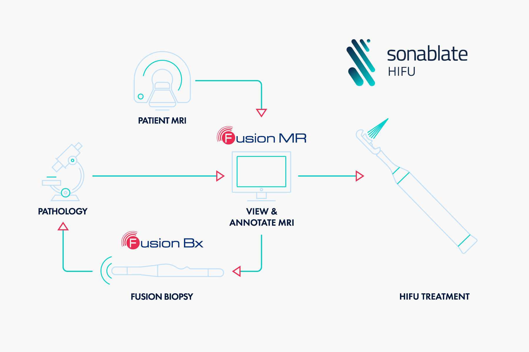
The primary checkpoint to undergo focal therapy is a positive fusion biopsy. Fusion biopsies give a more detailed picture of the patient’s histology, which directs the plan of action. They are drivers for treatment because they detect more, and often missed, clinically significant cancer. Just like targeted biopsies, focal therapy requires two things: an MRI and fusion. First a patient undergoes an MRI, which is read and annotated using Fusion MR Viewer. The output is accessed by the Fusion Bx and then a fusion biopsy is performed to confirm the cancer. Once pathology confirms positive results, the output (series of MR images) from Fusion MR Viewer is imported into Sonablate HIFU. Sonablate HIFU fuses these images with its ultrasound, which allows the urologist to plan the treatment based on the MRI and positive biopsy results. By adopting both the Fusion Bx and Sonablate HIFU, physicians can create targeted biopsy and treatment plans based on patients’ needs. For more information on Sonablate HIFU and Fusion Bx compatibility contact us at [email protected].
Sonablate Corp. (n.d.). What is HIFU? Retrieved from https://www.sonablate.com/products-procedures
Barkin, Jack. (2011). High intensity focused Ultrasound (HIFU). The Canadian Journal of Urology, 18(2), 5634-43.
Illing, R., & Emberton, M. (2006). Sonablate®-500: Transrectal high-intensity focused ultrasound for the treatment of prostate cancer. Expert Review of Medical Devices, 3(6), 717-729. doi:10.1586/17434440.3.6.717